Bone Erosion Research Tophi create an imbalance in the bone remodeling process that can cause bone erosions1-3

Over the last decade, Professor Nicola Dalbeth, MD, has published multiple papers featuring her industry-leading research on bone erosion and joint damage from gout. Using advanced imaging techniques like dual-energy computed tomography scans, Professor Dalbeth’s research has shown a clear correlation between urate deposition and bone erosion.4
The correlation between tophi and bone erosion in gout
with Professor Nicola Dalbeth, MD
Dr Dalbeth: Flares are not the only aspect of the disease. And tophi, even if they’re not acutely inflamed and causing severe pain at that very moment, have a number of really important consequences for patients. And I think often as clinicians that’s more difficult to actually appreciate if we don’t really start examining and looking at those sites.
We know that bone erosion is a consequence of long-standing gout. We do see this quite often in people who have tophaceous disease. But we really didn’t have a sense at that time what was actually contributing to the joint damage that we often saw in patients with long-standing, severe disease.
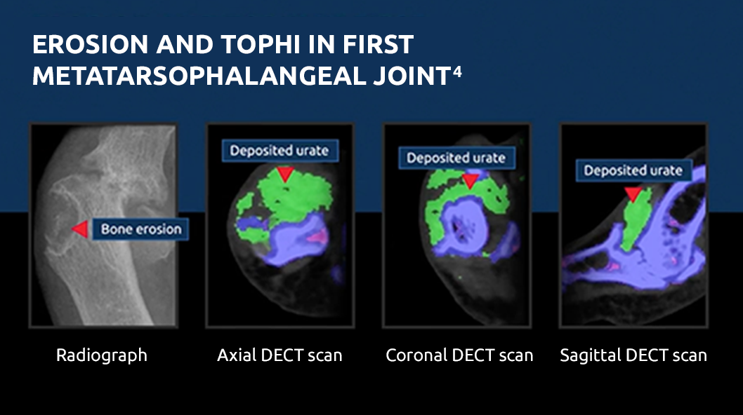
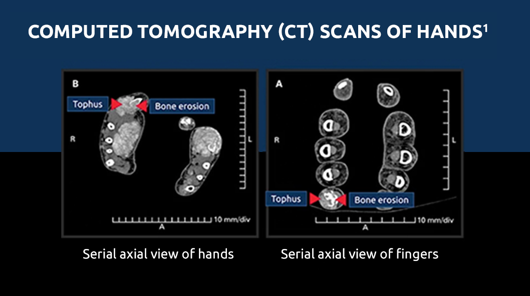
Images have been adapted with permission from Dalbeth N et al. Ann Rheum Dis. 2015;74:1030-1036.4 Watch at 00:47
And what we saw very clearly was that those joints, where there was definite and clear bone erosion, in those joints, there was virtually always evidence of a tophus present as well.
We also looked and compared the size of the erosion and the size of the intraosseous tophus, and again demonstrated a very close relationship between those 2 structures.
We were very interested in trying to understand the mechanisms of what was actually causing bone erosion.
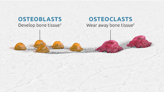
In the healthy skeleton, we see that this is really a dynamic, constant process of essentially replacing old or fatigued bone with new bone. Osteoclasts, osteoblasts—these cells talk to each other, they communicate, they work, in a normal skeleton, in unison.
This is quite a complicated, dynamic system, but certainly we really do see all of the cells that are involved in bone remodeling affected at these sites of erosion.
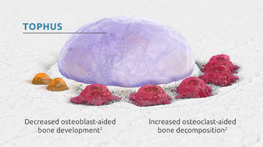
MSU crystals have direct effects on osteoblasts to reduce osteoblast viability.
If we look at that interface, so that tophus bone erosion interface, we see very few osteoblast cells lining the bone. So, essentially there’s reduced bone formation at these sites.
We see accelerated osteoclastogenesis. There’s a number of different mediators within the tophus that promote osteoclastogenesis.
This really provides further data to support the concept that actually the tophus within the joint is really driving structural damage.
Images adapted with permission from Dalbeth N. Ann Rheum Dis. 2009;68(8):1290-1295.1 Watch at 02:36
I don’t think we have a good concept of actually how rapidly erosion can occur in patients with tophaceous disease. Often when we’re seeing patients, you know they’ve developed tophi, and when we x-ray them, they already have established erosions.
I think it does give me pause to think that actually this is a serious complication of gout. And that we should be taking tophaceous disease seriously.
I think that that really does lend urgency to the importance of getting the serum urate down to low enough levels to be able to actually dissolve the crystals.
Learn more about how
gout affects the joints